Ehlers-Danlos syndromes (EDS) are a range of 14 disorders that primarily affect connective tissues, causing them to become less structurally sound. This manifests in a variety of localities, including the skin, ligaments, joints, internal organs, and blood vessels. The most well-known symptom of EDS is abnormally stretchy skin, but other symptoms can include joint hypermobility, extensive bruising, and skeletal dysplasia depending on which specific disorder a patient has. The spondylocheirodysplastic form of EDS (SCD-EDS) is characterized by a short stature, muscle hypotonia, and shortening and weakening of the bones, in addition to the classical symptoms of EDS. (Malfait & de Paepe, 2014)
The symptoms of all forms of EDS are ultimately caused by an alteration to the normal structure and function of collagen. Collagen is the most common structural extracellular matrix protein and is found in large quantities in connective tissues. In the case of EDS, genetic mutations on three different levels can cause these alterations (Malek & Köster, 2021). The first level of mutations are mutations directly to collagen genes, as seen in the classical form of EDS. These mutated genes encode collagen proteins that are misshapen, which impairs their function. The second level of mutations are found in genes that encode proteins which modify the collagen proteins after their translation. An example of this is found in kyphoscoliotic EDS, where a mutation to the PLOD1 gene results in a dysfunctional lysyl hydroxylase protein. Lysyl hydroxylase converts lysine residues on collagen fibers to hydroxylysine. This process is essential for proper structure of the collagen network, and its dysfunction causes the symptoms of this form of EDS (Xiao & Zhao, 2018). The last level of mutations are to genes that encode proteins that interact with the proteins that modify collagen; these indirect mutations still cause many of the same symptoms found in classical EDS. One such mutation has been found to cause SCD-EDS, where a mutation to the SLC39A13 produces a dysfunctional ZIP13 protein. There are various models that attempt to explain how this mutation causes SCD-EDS, but all models agree that a dysfunctional ZIP13 protein impairs the activity of lysyl hydroxylase, thereby creating an abundance of deformed collagen fibers (Xiao & Zhao, 2018).
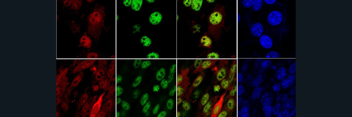
The first of such models devised to explain the pathological mechanism of SCD-EDS was based on the research conducted by Fukada et al. in 2008. These researchers observed that mice with a SLC39A13 knockout displayed similar symptoms to human patients with SCD-EDS, namely delayed growth, spine curvature, and bone abnormalities like osteopenia. They also saw a decrease in collagen volume and density in the bones and the dermal layers of the skin. This confirmed that the mutations in the SLC39A13 gene that produced a dysfunctional ZIP13 protein were responsible for the symptoms of SCD-EDS, but the exact mechanism was still unknown. To further investigate this, the researchers explored where the ZIP13 transporter was localized to within the connective tissue cells. Proteins in the ZIP family are known zinc transporters, and they all are found on either the outer cell membrane or on the membranes of intracellular compartments (Bin et al., 2014). It was found that the ZIP13 transporter localized specifically to the Golgi apparatus, not the outer membrane or on other intracellular compartments.
The paper published by Fukada et al. in 2008 helped to lay the foundation for SCD-EDS research, and helped other researchers describe the first mechanistic model for this disorder. This first model postulated that the lack of ZIP13 on the membrane of the Golgi leads to a buildup of zinc in this compartment, as the transport activity of the protein does not occur. The excess zinc in the Golgi then competes with iron for a binding site on lysyl hydroxylase, which is active in the Golgi and requires iron to be bound for its proper function. Lysyl hydroxylase activity would then be inhibited, meaning it cannot modify collagen, resulting in deformed collagen (Xiao & Zhao, 2018). It was thought that this pathway is what ultimately caused the symptoms of SCD-EDS, although two other models have emerged since then. I recommend the paper by Xiao & Zhao published in 2018 for further information on this interesting story.
References:
Bin, B. H., Hojyo, S., Hosaka, T., Bhin, J., Kano, H., Miyai, T., … & Fukada, T. (2014). Molecular pathogenesis of Spondylocheirodysplastic Ehlers‐Danlos syndrome caused by mutant ZIP13 proteins. EMBO molecular medicine, 6(8), 1028-1042.
Fukada, T., Civic, N., Furuichi, T., Shimoda, S., Mishima, K., Higashiyama, H., … & Hirano, T. (2008). The zinc transporter SLC39A13/ZIP13 is required for connective tissue development; its involvement in BMP/TGF-β signaling pathways. PloS one, 3(11), e3642.
Malek, S., & Köster, D. V. (2021). The Role of Cell Adhesion and Cytoskeleton Dynamics in the Pathogenesis of the Ehlers-Danlos Syndromes and Hypermobility Spectrum Disorders. Frontiers in Cell and Developmental Biology, 9, 883.
Malfait, F., & Paepe, A. D. (2014). The ehlers-danlos syndrome. Progress in heritable soft connective tissue diseases, 129-143.
Xiao, G., & Zhou, B. (2018). ZIP13: a study of Drosophila offers an alternative explanation for the corresponding human disease. Frontiers in genetics, 8, 234.